Imaging
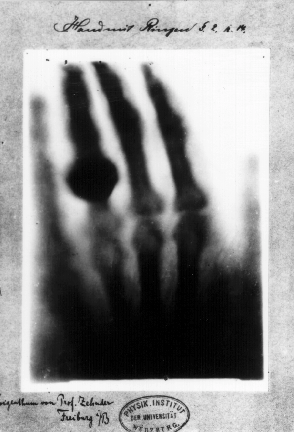
Imaging is the first historic application using X-rays, as demonstrated already by W. C. Röntgen, and remains the most common application especially due to its wide use for medical imaging.
Since the first X-ray image in 1895, an enormous development of X-ray equipment has taken place. Even though most imaging done today uses the same method as Röntgen did, the image quality has become far better thanks to the improved sources and detectors. Nowadays, X-ray imaging is also widely used in various fields, from industrial inspection & metrology to academic research.
The improved equipment has also opened up the possibility of new methods for X-ray imaging. They all differ in imaging performance and in their requirements on the equipment. Below we give a brief overview of popular imaging methods together with application examples using the MetalJet and NanoTube sources.
All the methods can be used either in two-dimensional projection imaging or three-dimensional computed tomography (CT). Whichever to use depends on the specific needs in the application.
